Part 1: Average expression level of specific gene across cell type
Download expression matrix of all expressed genes across cell types
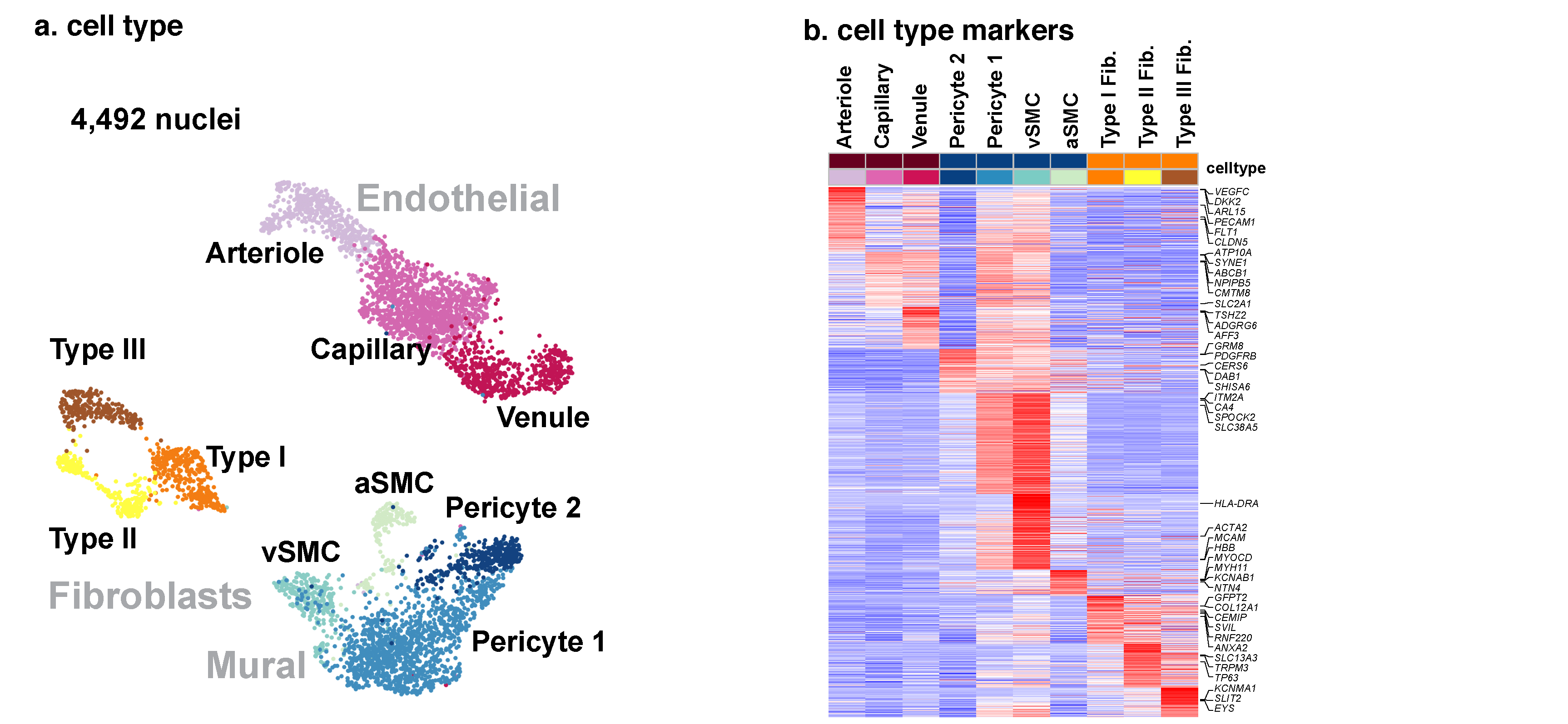
Part 2: Cell type and markers
Despite the importance of the cerebrovasculature in maintaining normal brain
physiology and in understanding neurodegeneration and drug delivery to the central
nervous system1, human cerebrovascular cells remain poorly characterized owing to
their sparsity and dispersion. Here we perform single-cell characterization of the
human cerebrovasculature using both ex vivo fresh tissue experimental enrichment
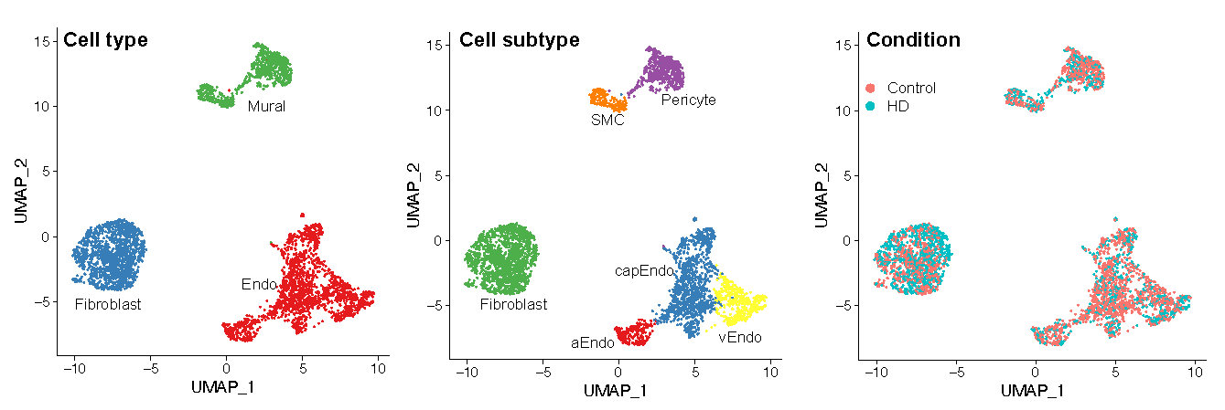
and post mortem in silico sorting of human cortical tissue samples. We capture 16,681
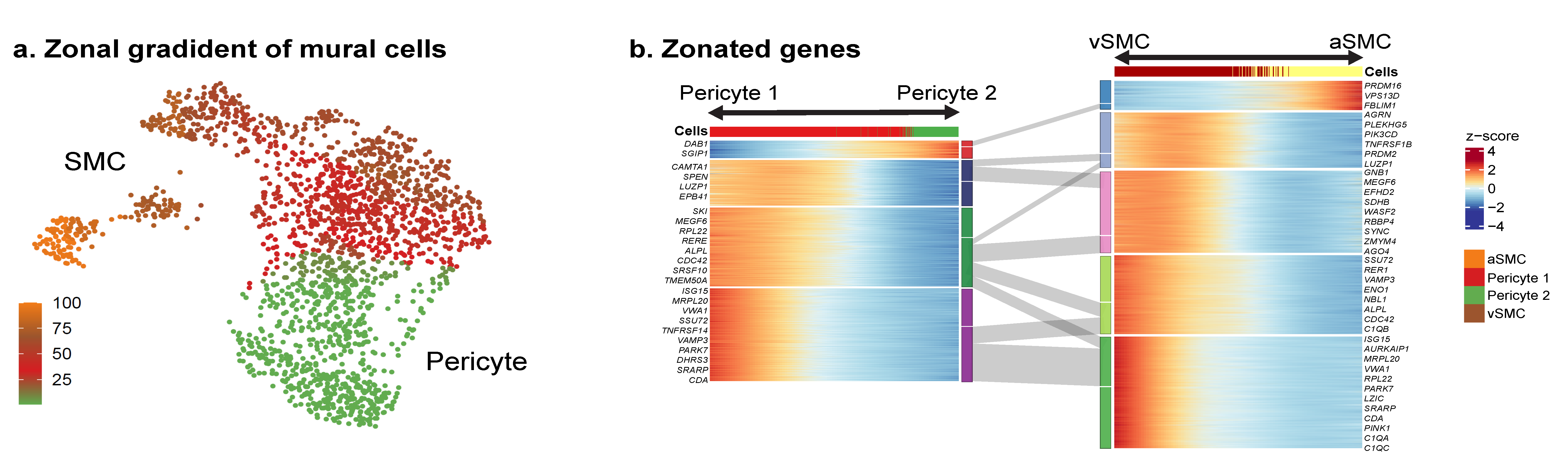
cerebrovascular nuclei across 11 subtypes, including endothelial cells, mural cells and
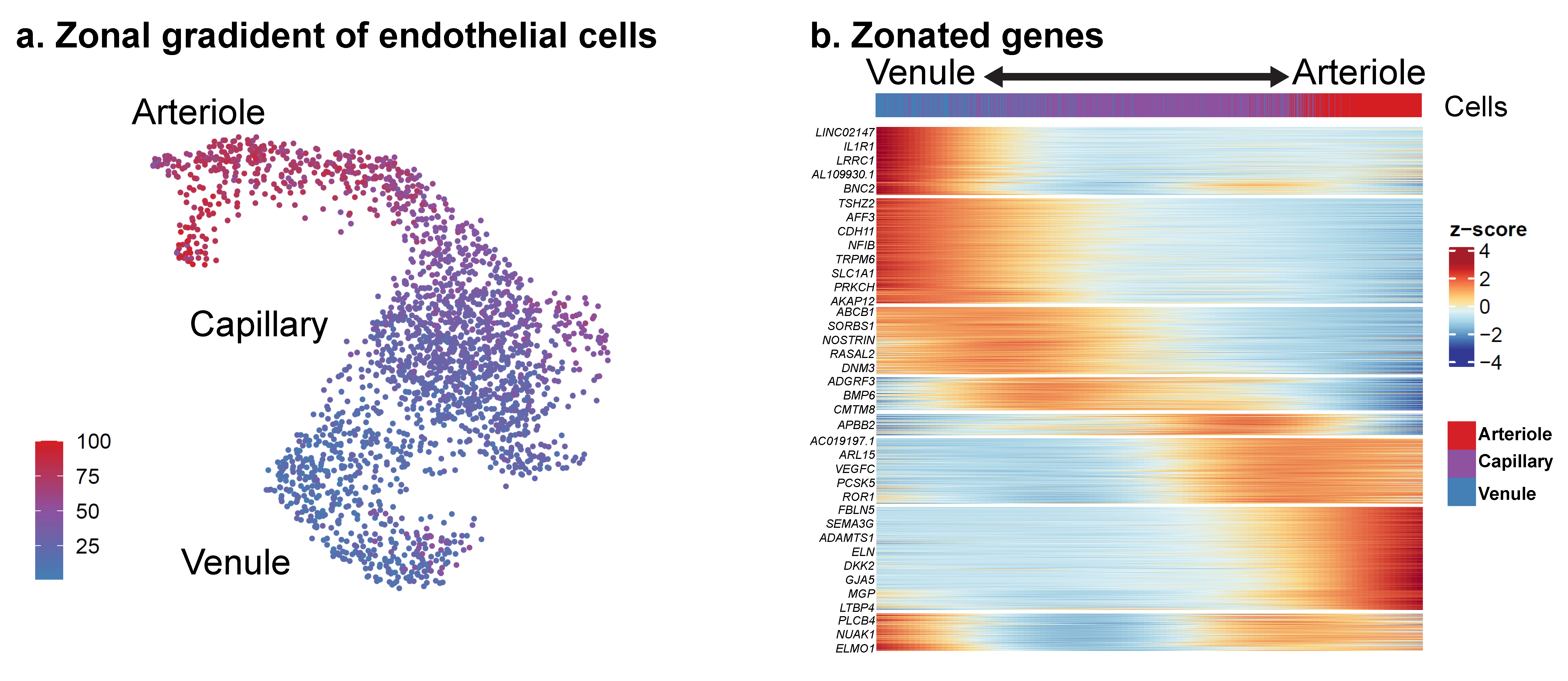
three distinct subtypes of perivascular fibroblast along the vasculature. We uncover
human-specific expression patterns along the arteriovenous axis and determine
previously uncharacterized cell-type-specific markers. We use these human-specific
signatures to study changes in 3,945 cerebrovascular cells from patients with
Huntington’s disease, which reveal activation of innate immune signalling in vascular
and glial cell types and a concomitant reduction in the levels of proteins critical for
maintenance of blood–brain barrier integrity. Finally, our study provides a
comprehensive molecular atlas of the human cerebrovasculature to guide future
biological and therapeutic studies.
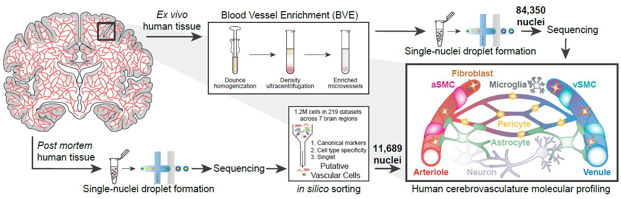
Figure 1 Overview